Technological frontier
How to prevent enterosporidiosis
Today, there are two main culprits threatening shrimp farming, one is early mortality syndrome (EMS) caused by infection with Vibrio parahaemolyticus; big. At present, there are many prevention and treatment methods for EMS, but there is no effective treatment method for EHP so far. Affected by the disease that has persisted in successive years, the confidence of shrimp farmers has been damaged, and the farming area and output have been declining.
I. Pathogens of Microsporidia
The causative agent of microsporidia is Microsporidia, which is a single-celled eukaryotic organism that parasitizes intracellularly. , fur animals and humans, but mainly parasitic in invertebrates such as shrimps and crabs. There are many species of microsporidia, and most of the microsporidia parasitic on shrimp belong to four genera in the family Microsporiaceae, namely Octosporidia, Microsporidia, Piriformis and EHP. The main parasitic site of Microsporidia of the genus Microsporidia is the muscle tissue of prawns. Piriformis not only parasitizes muscle tissue, but also infects the heart, gills, stomach, liver and intestine. Intestinal epithelial cell Microsporidia parasites in the intestine and liver pancreas.
two. Transmission Routes of Microsporidia Diseases
After the spores are swallowed into the digestive tract by shrimps and crabs, they are rapidly activated and then continue to infect the intestines, liver or other tissues. The infection route of Microsporidia is mainly from the mouth into the digestive tract to infect shrimp and crab cells. Therefore, controlling the feed, water quality and environment from being infected by microsporidia is the main way to prevent and treat microsporidia. However, there is no intergenerational transmission of microsporidiosis, and broodstock should not directly transmit microsporidiosis to shrimp fry.
Three. Symptoms of Microsporidia Disease
Symptoms of microsporidia infection with microsporidia in prawns mainly include the following:
1. Shrimp
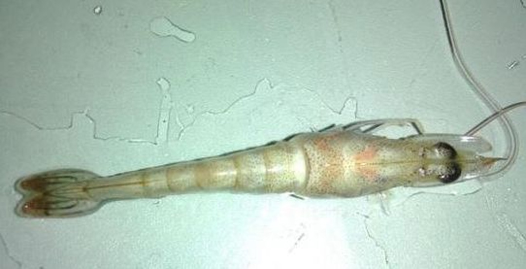
The body of the shrimp infected with microsporidiosis turned white, and after peeling off the carapace, the muscle tissue was milky white. However, it must be distinguished from the symptoms caused by excessive nitrite. When the body of the shrimp is found to be cloudy, the possibility of nitrite poisoning must be ruled out first, so as to avoid irreparable losses caused by delaying the rescue time.
2. Cotton Shrimp
It mainly infects the gonads and striated muscles of prawns, causing the muscles to be cloudy, opaque, and lose elasticity, which is called milky white shrimp or cotton shrimp. If cotton shrimp appears in diseased shrimp infected with microsporidiosis, there is basically no possibility of recovery. If it is cotton shrimp caused by malnutrition, supplementation with appropriate amino acids or high-quality animal protein can significantly improve the effect in about 3 days. The appearance of such cotton shrimp can be recovered.
3、Not grown up
After shrimp infection with Microsporidia EHP in intestinal epithelial cells, the most obvious feature is that the growth rate becomes slow, resulting in large and uneven individual differences in shrimp. However, shrimp infected with EHP will not die immediately. Their intestines are also very full, their appetite is normal, and they can eat normally. On the surface, shrimps do not have obvious symptoms of disease, but they do not grow long after eating only, and occasionally white stools are accompanied, causing huge economic losses.
IV. Microsporidia EHP in intestinal epithelial cells
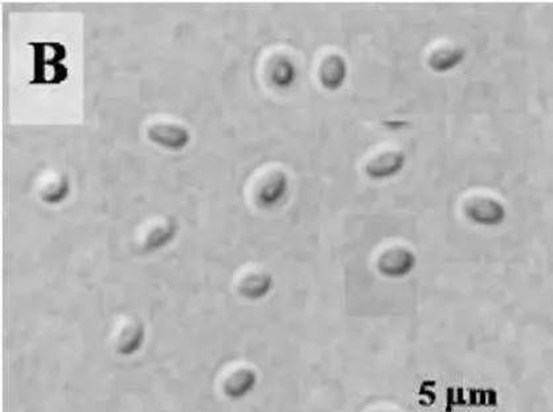
Intestinal Epithelial Microsporidia (referred to as Enterosporidium, EHP) is caused by a microsporidian parasite, named in 2009, after symptoms were found in slow-growing Penaeus monodon in Thailand, EHP is limited to Shrimp gut and hepatopancreas. Enterosporidium is a small sporozoite with a body length of less than 1 micron. It mainly infects the intestinal epidermis and hepatopancreas of prawns, resulting in a decrease in the intestinal absorption function of prawns, and severe enteritis and hepatopancreas atrophy. Enterosporidium develops from the early protoplasmic stage to mature oval spores in the cytoplasm of the hepatopancreas, with a size of 0.7 × 1.1 microns, a single nucleus, 5 to 6 circles of polar filaments, an anchor disc connecting the polar filaments, and a rear end with a Vacuole, sporophyte dense and hard. Enterosporidium can be transmitted vertically or horizontally, such as vertical transmission from seedlings carrying pathogens, or horizontal transmission by enterosporidium-contaminated breeding ponds.
Intestinal epithelial cell microspore electron microscope image below:
5. The difference between enterosporidiosis and infectious subcutaneous and hematopoietic necrosis virus disease
Infectious hypodermic and hematopoietic necrosis virus (IHHNV) can cause shrimp to suffer from chronic "short stature and disability syndrome", which affects the growth of shrimp, and also makes shrimp small. The carapace is rough, dull or incomplete and other lesions, which are manifested as inflammatory lesions of gastric epidermis, hindgut, gills, striated muscles, ganglia, connective tissues, blood cells, and hematopoietic organs, and severe necrosis. Shrimp infected with infectious hypodermic and hematopoietic organ necrosis virus carry the virus for life, which can be transmitted to other groups through mutual infection, the virus can also be transmitted to the next generation through fertilized eggs, and can also be transmitted through the same kind of residual food. When the environment of the aquaculture water body deteriorates, the diseased shrimp will be accompanied by a large number of deaths.
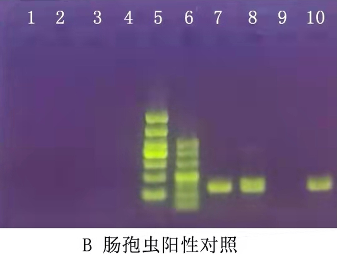
6. Diagnosis of Enterosporidiosis To diagnose whether shrimps are infected with Enterosporidiosis, laboratory nested PCR testing is needed to confirm the DNA.
The polymerase chain reaction (PCR) uses a piece of DNA of the sample to be tested as a template and a specific oligonucleotide sequence complementary to both ends of the template as a primer. DNA under the action of DNA polymerase. After dozens of reaction cycles of denaturation, annealing and extension, the DNA fragments between the two primers on the template are amplified in a specific series, and the specifically amplified fragments are detected by electrophoresis and other means. Thus, the presence of trace amounts of viral DNA can be revealed. The nested PCR used in this experiment is to use a pair of primers to carry out the second-step PCR amplification in the product of the first-step PCR amplification. Nested PCR can greatly increase the sensitivity and reaction specificity of PCR detection.
VII. Prevention and control measures of shrimp enterosporidiosis
At present, there are no effective measures for the treatment of enterosporidiosis in shrimp. Therefore, it is very important to do the following preventive measures:
1. Choose pathogen-free shrimp
Shrimp fry carrying any kind of pathogen are not put in. Before purchasing shrimp seedlings, first pass the PCR test to check whether the shrimp seedlings carry bacteria, viruses and sporozoites to ensure that the shrimp seedlings do not carry any pathogens.
2. Strictly control shrimp bait
Shrimp seedlings should be fed live bait and bait that is not infected with Enterosporidium, to prevent shrimp seedlings from being infected with Enterosporidium by eating bait with Enterosporidium.
3. Insecticide and disinfection of shrimp ponds
In shrimp ponds that were previously infected with Enterosporidium, the spores of Enterosporidium were in a dormant state and lurked in the soil of shrimp ponds. Chlorine, iodine, pesticides and other drugs are not easy to kill. At present, the strong alkali disinfection method is used to kill Enterosporidium spores in the soil. 200kg of quicklime is used per mu, withdrawn to the bottom of the dry pond, ploughed about 10 cm of the bottom mud, and added about 10 cm of water, the pH value of the soil will rise to above 12 Thereby killing Enterospora. However, it is exposed to the sun for more than half a month, or directly to the next year for breeding. This method can not only kill Enterosporidium, but also effectively kill wild fish, harmful organisms, parasites, pathogenic bacteria, etc. in the fish pond, and can also ferment the organic matter at the bottom of the pond into organic calcium fertilizer. Farmers do not have to worry that the use of a large amount of quicklime will cause the PH value of the shrimp aquaculture water to rise, because the quicklime water will absorb carbon dioxide and change it into calcium carbonate, so that the pH value will fall back to the normal range (10 days can be the pH value drops to normal). scope).
4. Enhance the immunity of prawns
The physique of prawns is strong, and the risk of contracting any pathogen is much lower. During the breeding period, it is recommended to feed high-grade shrimp compound feed with balanced nutrition, orally take health products such as multivitamins, small peptides, oligosaccharides, trace elements, etc., to enhance the disease resistance of shrimp, and feed as little fresh bait as possible to prevent unknown pathogens from entering the pool. lead to death of prawns.
R&D Center: Liu Fang
ghxiedaojia

|Guanghe crab rice home|
WeChat public account
Aquaculture knowledge
share with you

